
3-photon microscopy for deep-tissue imaging
Introduction
Imaging depth in two-photon (2P) microscopy is limited by scattering. At depths beyond 500 µm in a densely stained mouse brain, 2P excitation confinement and signal-to-background ratio are severely degraded. One workaround recently pioneered at Cornell University (Xu et al, Nat Phot 2013) is to use 3-photon excitation with infrared-shifted wavelengths near 1.3 or 1.7 µm. This approach can significantly improve the imaging depth, owing to superior excitation confinement (3P effect) and reduced scattering (infrared effect). 3P microscopy makes it possible to image cells at depths exceeding 1 mm in live mouse brain tissue. In addition, 3P fluorescence imaging is easily combined with morphological imaging based on third harmonic generation (THG). 3P microscopy recently became practical with the development of suitable high-peak power infrared sources. We are developing this promising approach and novel applications.
Dual-color 3-photon microscopy
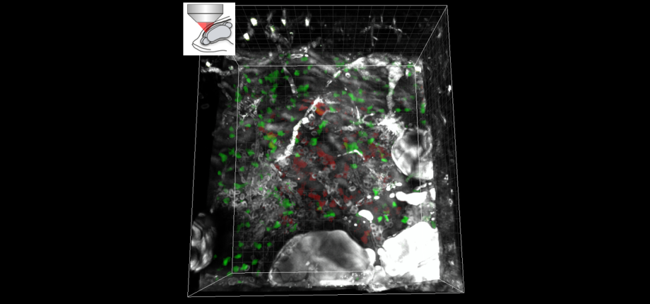
In collaboration with the 'Lasers' group at IOGS, we introduced a dual-color laser source design optimized for 3P excitation; we used it to build a 3-photon microscope capable of exciting simultaneously green (GFP) and red (RFP, dTomato) fluorescent proteins deep inside brain tissue (along with label-free THG signals), and demonstrated 3-photon through-skull images of live adult zebrafish brain.
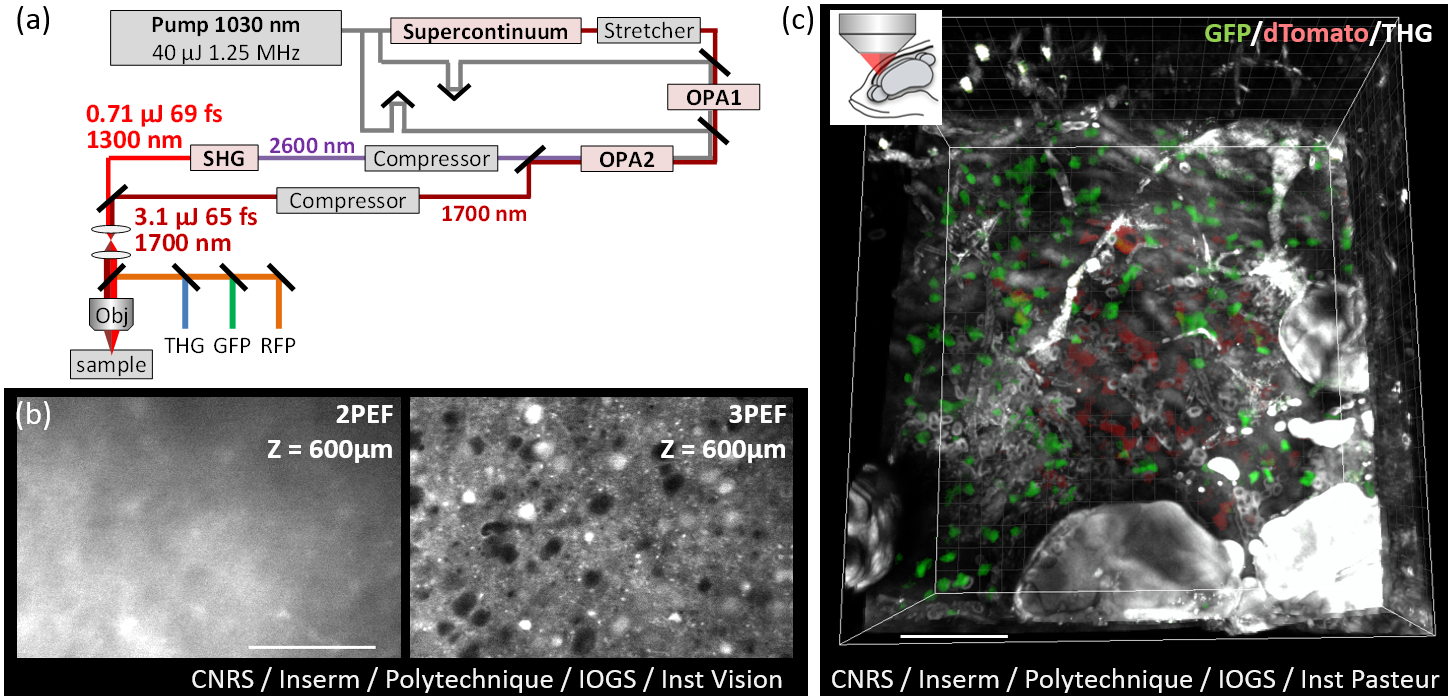
(a) Dual-color laser source optimized for 3P microscopy. (b) Comparison of 2P and 3P in-depth imaging of ex vivo mouse brain tissue. (c) in vivo through-skull 3P/THG imaging of two neural cell populations in an adult zebrafish brain. © CNRS, Inserm, Polytechnique, IOGS, Inst Vision, Inst Pasteur / Springer-Nature. Adapted from Guesmi, Abdeladim et al, Light Sci App 2018.
Publications
Label-free imaging of red blood cells and oxygenation with color third-order sum-frequency generation microscopy
J. Ferrer Ortas, P. Mahou, S. Escot, C. Stringari, N. B. David, L. Bally-Cuif, N. Dray, M. Négrerie, W. Supatto, E. Beaurepaire
Light: Science & Applications (2023)
Intravital deep-tumor single-beam 3-photon, 4-photon, and harmonic microscopy
G.-J. Bakker, S. Weischer, J. Ferrer Ortas, J. Heidelin, V. Andresen, M. Beutler, E. Beaurepaire, P. Friedl
eLife (2022)
Color imaging with multimodal 3-photon microscopy
Optics & Photonics News (2018)
Dual-color deep-tissue three-photon microscopy with a multiband infrared laser
K. Guesmi, L. Abdeladim, S. Tozer, P. Mahou, T. Kumamoto, K. Jurkus, P. Rigaud, K. Loulier, N. Dray, P. Georges, M. Hanna, J. Livet, W. Supatto, E. Beaurepaire, F. Druon
Light: Science & Applications (2018).
All-fiber femtosecond laser providing 9 nJ, 50 MHz pulses at 1650 nm for three-photon microscopy
P. Cadroas, L. Abdeladim, L. Kotov, M. Likhachev, D. Lipatov, D. Gaponov, A. Hideur, M. Tang, J. Livet, W. Supatto, E. Beaurepaire, S. Février
J. Optics (2017).