FLIM-based metabolic imaging

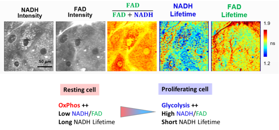
Fluorescence Lifetime Microscopy (FLIM) of intrinsic biomarkers has demonstrated an exceptional capability to non-invasively investigate the energetic and metabolic redox state of living tissues at the cellular level.
We develop novel approaches for fast FLIM imaging and label-free optical methods for metabolic imaging in stem cells and living tissues.
Contact: Chiara Stringari
Related publications
Label-free nonlinear microscopy probes cellular metabolism and myelin dynamics in live tissue.
Asadipour B, Morizet J, Ronzano R, Zhang X, Aigrot MS, Mahou P, Solinas X, Phan MS, Chessel A, Stankoff B, Desmazieres A, Beaurepaire E, Stringari C.
Commun Biol. (2025)
Consensus guidelines for cellular label-free optical metabolic imaging: ensuring accuracy and reproducibility in metabolic profiling
Georgakoudi I, Skala MC, Quinn KP, Stringari C, et al.
J Biomed Opt. (2025)
Emerging Functional Connections Between Metabolism and Epigenetic Remodeling in Neural Differentiation
E. Sánchez-Ramírez, TPL. Ung, C. Stringari, L. Aguilar-Arnal
Molecular Neurobiology (2024)
FLUTE: A Python GUI for interactive phasor analysis of FLIM data
D. Gottlieb, B. Asadipour, P. Kostina, T.P.L. Ung, C. Stringari
Biological Imaging (2023)
Label-free single-cell live imaging reveals fast metabolic switch in T lymphocytes
N. Paillon, TPL. Ung, S. Dogniaux, C. Stringari, C. Hivroz
Mol Biol Cell (2023)
Coordinated metabolic transitions and gene expression by NAD+ during adipogenesis
E. Sánchez-Ramírez, T.P.L. Ung, A. Alarcón del Carmen, X. del Toro-Ríos, G.R. Fajardo-Orduña, L.G. Noriega, V.A. Cortés-Morales, A.R. Tovar, J.J. Montesinos, R. Orozco-Solís, C. Stringari, L. Aguilar-Arnal, J Cell Biol (2022)
Simultaneous NAD(P)H and FAD fluorescence lifetime microscopy of long UVA–induced metabolic stress in reconstructed human skin
T.P.L. Ung, S. Lim, X. Solinas, P. Mahou, A. Chessel, C. Marionnet, T. Bornschlögl, E. Beaurepaire, F. Bernerd, A.-M. Pena, C. Stringari, Scientific Reports (2021)
Multicolor two-photon imaging of endogenous fluorophores in living tissues by wavelength mixing C. Stringari, L. Abdeladim, G. Malkinson, P. Mahou, X. Solinas, I. Lamarre, S. Brizion, J.-B. Galey, W. Supatto, R. Legouis, A.-M. Pena, E. Beaurepaire, Scientific Reports (2017)
In Vivo single-cell detection of metabolic oscillations in stem cells Stringari C, Wang H, Geyfman M, Crosignani V, Kumar V, Takahashi JS, Andersen B, Gratton E. Cell Rep (2015)
Metabolic trajectory associated with cellular proliferation in small intestine by Phasor Fluorescence Lifetime Microscopy of NADH Stringari C., Edwards RA, Pate KT, Waterman ML, Donovan PJ, Gratton E., Scientific Reports (2012)
Phasor approach to fluorescence lifetime microscopy distinguishes different metabolic states of germ cells in a live tissue. Stringari C., Cinquin A., Cinquin O., Digman MA., Donovan P., Gratton E. PNAS (2011)


